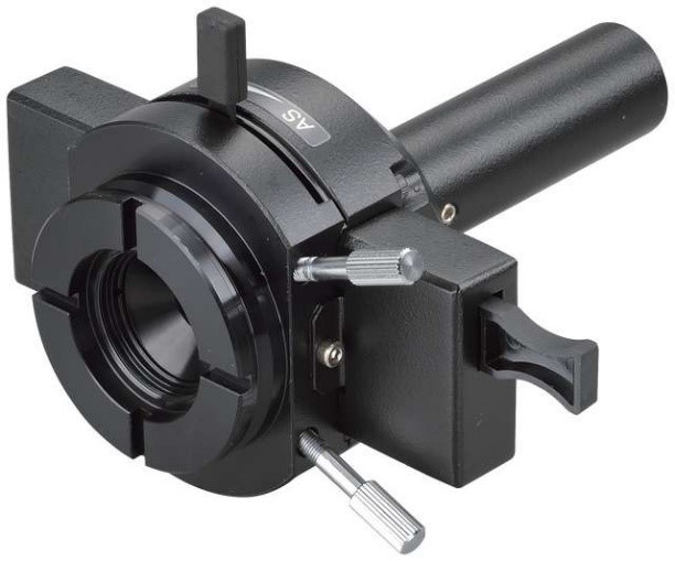
Nikon Ti2 - LAPP 模組
超越極限的顯微光學應用
• 多功能光學操作、模組彈性運用、展現極致的擴充性
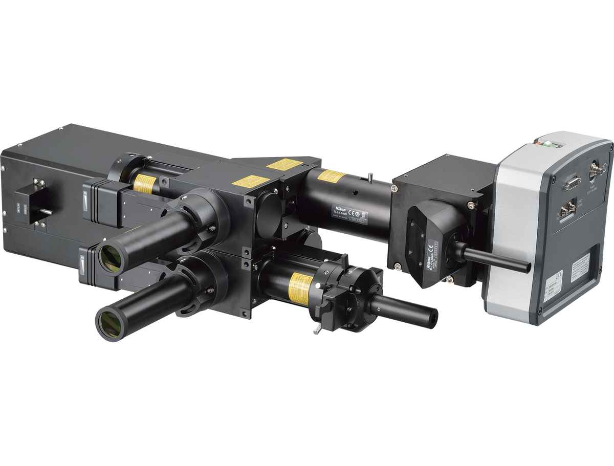
Nikon 的 Lapp 系列,將顯微光學裝置模組化,可依您的研究需求,選擇適當的光學模組,進行例如: Photoactivation / Conversion、Photobleaching 以及 TIRF 等實驗。目前這類應用已發表於多項期刊中。Lapp 目前可搭載於 Nikon 倒立顯微鏡 Eclipse Ti2 系列,模組式的組合最多可同時搭載五組不同的光操作模組(例如: 一組電動 TIRF、一組 N - STORM、一組 DMD、一組 FRAP 及一組 Epi - FL 模組)。
H - TIRF 模組
• 全自動光學校正 TIRF,讓您每次實驗都能達到最精準的觀測深度。
TIRF 為全內反射的光學應用模組,搭載 Nikon Eclipse Ti2 倒立式顯微鏡,可觀察液體介面下貼附於玻片表面約 200nm 深度之內的物質,是一種單分子顯微觀察方式。Nikon 的 H-TIRF可全自動光學校正,簡單上手,不須手動校正光源,輕鬆體驗 TIRF 帶來的震撼效果!
N - STORM 模組
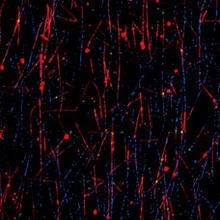
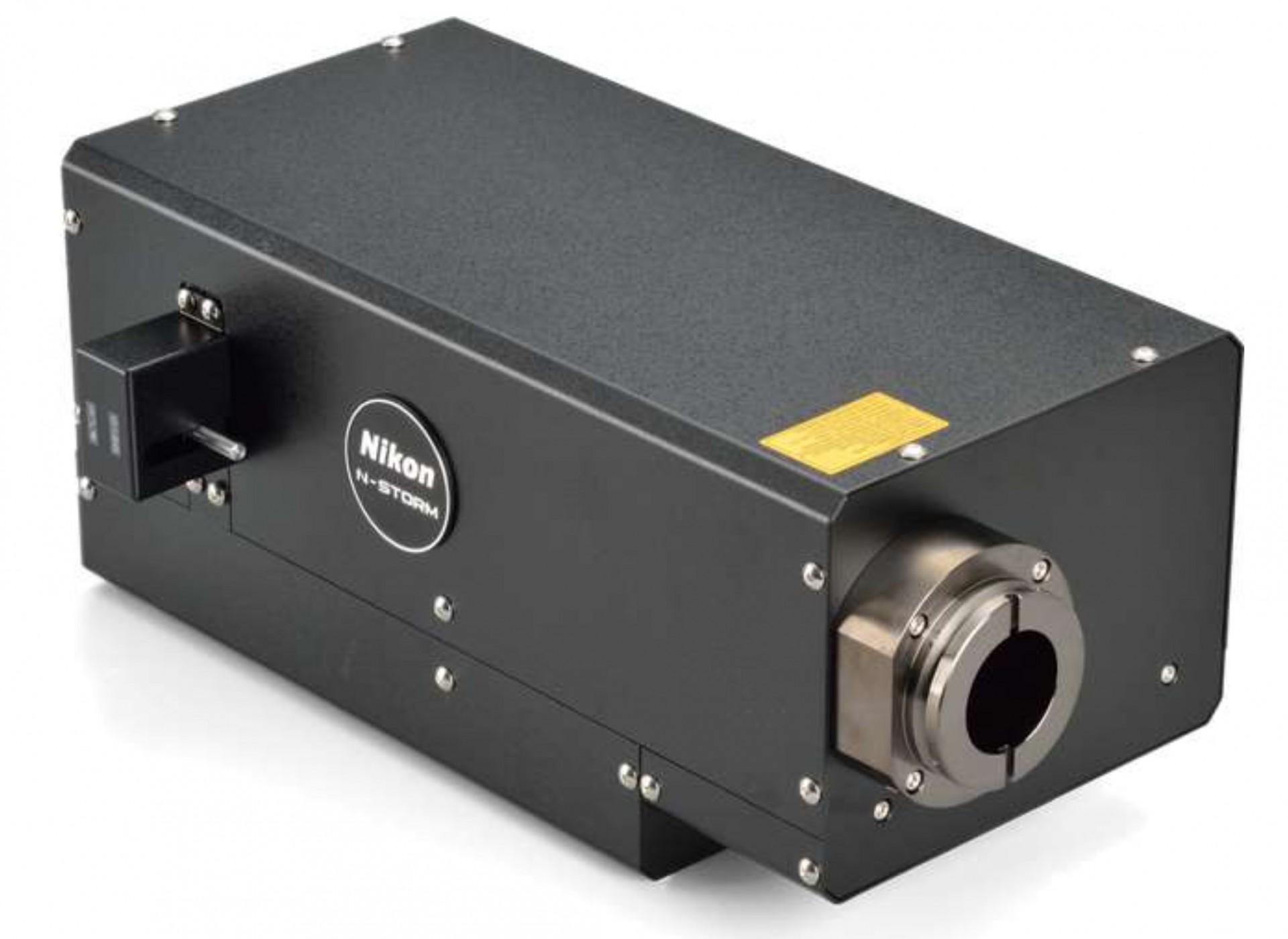
XY 軸光學分辨率可達 20nm,比傳統光學極限 200nm,提升 10 倍解析力,讓光學顯微鏡觀察範圍推向分子層級,開啟科學研究的新視野。
FRAP 模組
• 於繞射極限進行光操作實驗
FRAP 模組可於針對約 500nm 大小的單點來進行 Photoactivation / Conversion、Photobleaching 等光操作實驗,可搭配高 Frame - Rate 及高感光度的影像系統或是轉盤式共軛焦系統,是個經濟又有效率的裝置。
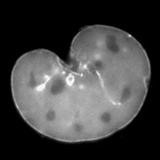
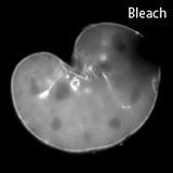
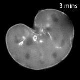
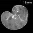
A mouse embryonic fibroblast expressing mCherry-lamin A was spot-photobleached in the upper right corner of the nucleus using the FRAP module to study the dynamics of lamin A molecules. Time-lapse images were acquired using the epi-fluorescence illuminator.
Image courtesy of Drs. Takeshi Shimi and Bob Goldman, Northwestern University Medical School
DMD模組
• 可同時多點進行光啟動(Photoactivation)實驗
有別於 FRAP 模組,DMD 模組可同時針對不同目標進行不同頻率的光活化實驗,尤其在 optogenetics 實驗,針對指定區域做光刺激後,觀察細胞中物質或蛋白質分布的變化。可選擇搭載不同類型的光源: LED 光源或 Laser 光源。
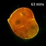
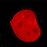
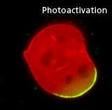
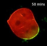
A mouse embryonic fibroblast co-expressing mCherry-tagged lamin A (red) and photo-activatable GFP-tagged lamin A was photo-converted (green) in the lower right region using the DMD module and 405 nm LED light. Time-lapse images were captured using the epi-fluorescence illuminator. By photoactivating a sub-population of the lamin proteins, one can observe their dynamics and subunit-exchange behavior. Image ourtesy of Drs. Takeshi Shimi and Bob Goldman, Northwestern University Medical School
XY 振鏡掃描模組
• 在共軛焦系統上,可同時進行成像與光刺激
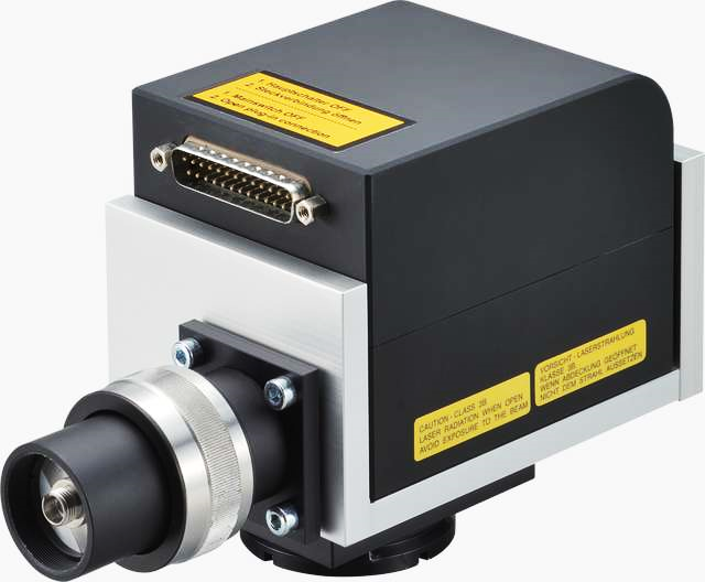
*本裝置可與 A1 HD25/A1R HD25搭配使用.
廣視野螢光照明模組
• 大面積的感應器不再無用武之地
