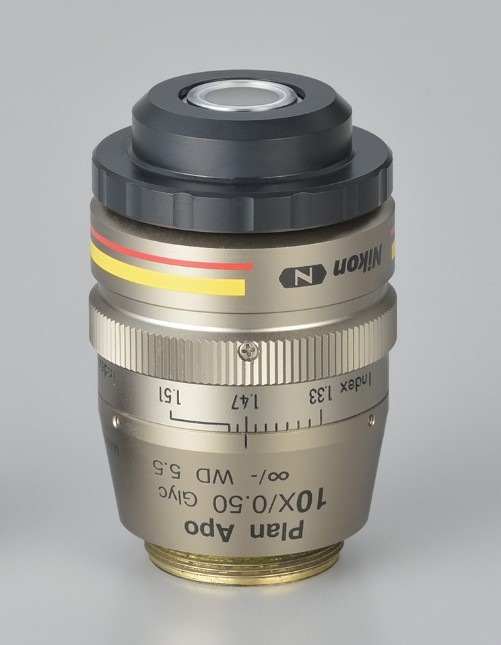
Plan Apochromat 10X Glyc 甘油鏡
Nikon 發表新的 10 倍 Plan APO 級的甘油鏡,此款甘油鏡的開發是為了處理厚樣本(deep tissue)的影像,適用於各式各樣的組織透明技術(tissue clearing technique),它的高折射率相當符合神經科學研究的需求。

開發動機
新的這顆 Nikon CFI Plan Apochromat 10 倍甘油鏡可以讓以往比較不易觀察的深層組織看起來就像清楚的大腦樣本一樣清晰。這顆新款的物鏡不僅提供了影像的高解析力(high resolution),更擁有大視野、高景深、高通量(high throughput)效果可應用在厚樣本的影像處理上(deep - imaging)。
主要特色
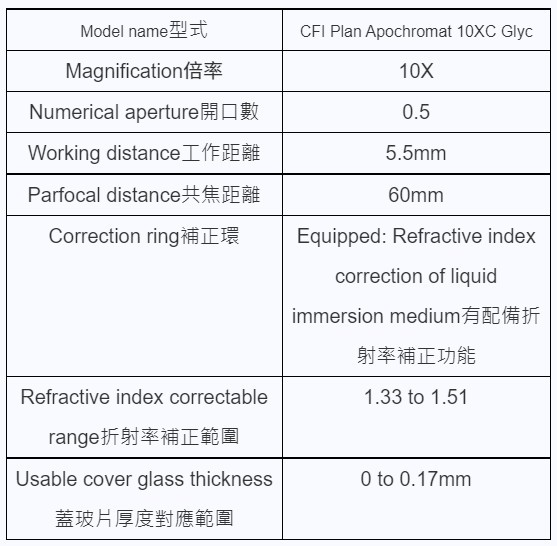
• 可調整對應折射率:具備校正環,可調整折射率範圍為 1.33 到 1.51,也就是說這顆物鏡具備了水鏡、油鏡、甘油鏡的功能,一鏡多用。
• 高穿透率:採用 Nikon 獨家奈米結晶鍍膜技術,UV 到近紅外光(near IR)的波長均有高穿透率。
• 消色差處理:由 405nm 到 1064nm,大範圍的消色差處理。對多光子顯微鏡拍攝厚樣本,尤其是腦組織,這類需要較長波長的實驗而言,是相當理想的工具。
參考照片

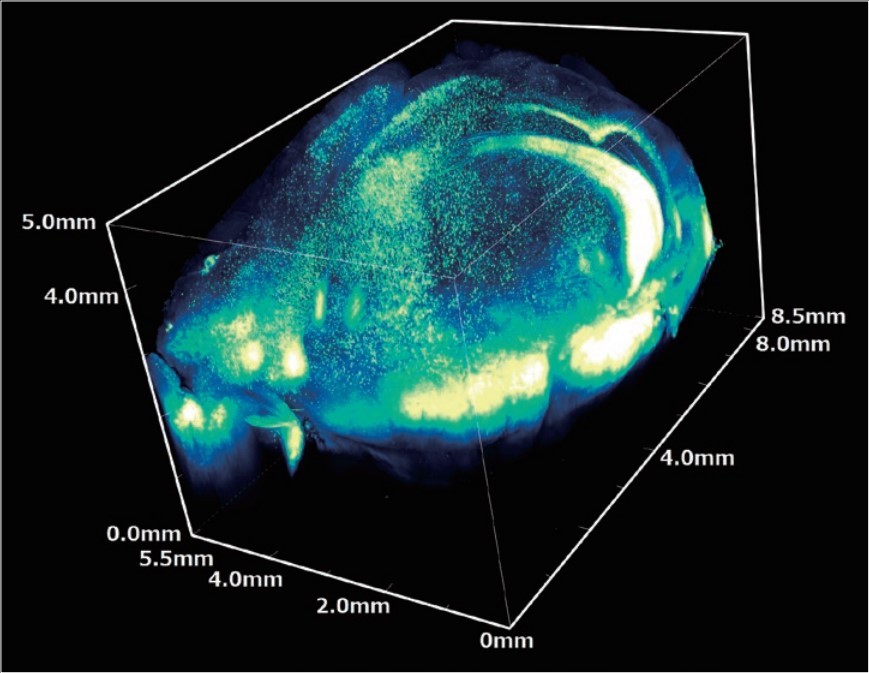
Fixed YFP-H mouse brain clarified with optical clearing solution.
*Stitched image captured with Nikon Multi Photon Microscope A1R MP+ , CFI Plan Apochromat 10XC Glyc.
Photographed with the cooperation of: Dr. Ryosuke Kawakami, Dr. Tomomi Nemoto, Research Institute for Electronic Science, Hokkaido University.
* Whole brain image with captured with transgenic mouse labeled with yellow fluorescent protein (YFP-H).
參考影像

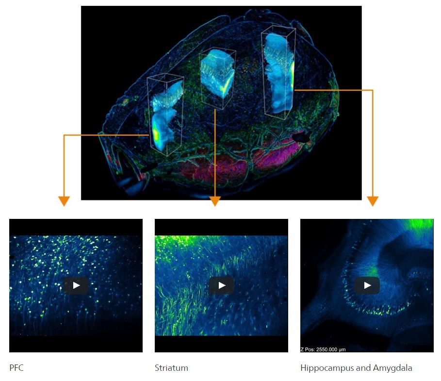
Optical slices of H-line mouse whole brain after clearing with LUCID-A.
Objective: CFI Plan Apochromat 10xC Glyc (N.A. 0.5, W.D. 5.5mm)
.Photo courtesy of: Drs. Ryosuke Kawakami and Tomomi Nemoto, Research institute for Electronic Science, Hokkaido University.
產品規格


